Macroscope: sensitive fluorescence imaging of whole zebrafish larvae
Quick facts
Summary
A 60 MP camera samples the specimen at 0.5 µm per pixel. Collecting 12× more light than a stereo microscope, it needs much shorter exposures for the same image quality. This sensitivity means you can place the instrument in a normally lit laboratory, no dark room needed.
A whole five day old zebrafish larva fits in one frame, with no zooming, tiling or stage moves, and live fluorescence at 10 fps keeps weak signals visible while you handle and inject samples. Also included: an overview camera, dome and telecentric lighting for brightfield, and a 4K OLED touchscreen.
Pre-ordering is available now for delivery in 2027. To arrange a demonstration or pre-order, sign up for the early access programme.
Low magnification is light-starved in current microscopes
Low-magnification fluorescence microscopy is often not sensitive enough to detect weak signals in whole organisms such as zebrafish larvae. The two standard instruments for this task each give up light in a different way:
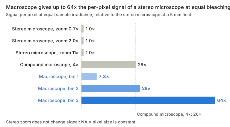
Stereo microscopes have a large objective lens that could collect a lot of light, but when zoomed out to see a whole larva (a 5 mm field), only about 3% of the lens area is actually used. This effectively reduces the objective’s NA from 0.57 to about 0.10. Zooming in does not help: you see a smaller area, but each camera pixel still receives the same amount of light.
Our example is a high-end fluorescence zoom microscope, among the most light-efficient of its kind; most stereo microscopes collect even less light.
Upright or inverted compound microscopes with a 4× objective collect about 4× more light than the stereo microscope, but that is still limited. A standard camera covers only about 3.3 mm, with fairly coarse pixels, which limits the detail you can see. Higher magnification means precise sample alignment, a motorised stage and stitching images together.
How old is the fluorescence stereo microscope in your lab, and how much of its light does it actually collect?
Optical design
The macroscope uses large-diameter optics: a 7×/0.35 NA objective with a Ø25 mm pupil feeding a tube lens and a 60 MP camera. This keeps the full aperture in use over the whole 4.5 × 3 mm² field, instead of stopping down to reach a low magnification.
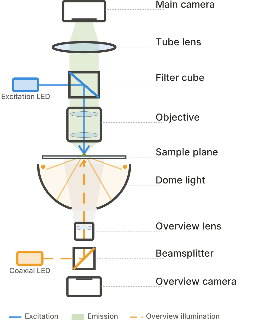
| Subsystem | Function |
|---|---|
| Main camera | 60 MP sensor, 0.5 × 0.5 µm² per pixel at the sample; 2×2 or 3×3 binning. Motorized focus, automated z-stack acquisition. |
| Fluorescence path | Excitation LED and large Ø50 mm filter cube, automated filter stage. |
| Sample plane | Accepts multi-well plates and petri dishes. |
| Dome light | Diffuse LED ring below the sample for high-NA brightfield transmission imaging. |
| Overview camera | Looks up through the dome for sample navigation and placement. |
| Coaxial LED | Coupled in via a beamsplitter through the overview lens; low-NA light that increases depth of field when imaging from above. |
Hardware binning increases both sensitivity and frame rate, enabling video-rate viewing of weak signals during sample handling.
Light collection at a matched 5 mm field
| Instrument | Magnification / NA | Pupil | Étendue (mm²·sr) | Photons (rel. to stereo) | Diffraction limit (µm) |
|---|---|---|---|---|---|
| Macroscope | 7× / 0.35 | Ø25 mm | 7.6 | 12× | 0.8 |
| Compound microscope, 4× | 4× / 0.20 | Ø20 mm | 2.5 | 3.9× | 1.4 |
| Stereo microscope, zoom 2.0× | 2.3× / 0.57 objective, 0.10 effective | Ø6.3 of Ø35 mm | 0.64 | 1.0× | 2.7 |
Signal per pixel at equal sample irradiance
Applications
The macroscope targets the manual bench work now done on fluorescence stereo microscopes, adding sensitivity and z-stack acquisition at the whole-embryo field. It can also replace a confocal system where that is used only for its sensitivity, not for sub-cellular resolution.
- Screening and sorting fluorescent embryos and larvae, including weak reporter lines that are hard to see without zooming in.
- Checking injections and xenografts: confirm that injected cells, dyes or particles reached their target.
- Whole-larva cell counting in infection, inflammation and xenograft models, such as GFP-labelled macrophages in MPEG-GFP larvae.
- Prescreening before confocal or light-sheet imaging, so expensive instrument time goes to the right samples.
- Publication-quality images of whole larvae: a stereo microscope is rarely used for figures, but the macroscope can quickly create image stacks from the whole larva at 0.5 µm pixels, in fluorescence and brightfield, without tiling or stitching.
- Z-stacks of the whole larva: at NA 0.35 the depth of field is only a few micrometres, so stepping focus through a larva brings successive planes into sharp focus across the full field. Software combines them into an all-in-focus (extended depth of field) image or a 3D stack. As with any widefield system, out-of-focus light is not rejected, so deconvolution helps for thick samples. A stereo microscope cannot do this at this field of view.
- Teaching and demonstration on touchscreen or external screen, with video-rate live viewing.
Optional modules offer more applications
- Heart rate and calcium imaging: 55 fps over the full field at 3×3 binning, or an estimated 75–90 fps on a brain- or heart-sized region with the large-field camera option.
- Fluorescence guided microinjection: with the injection module, injection can be done during video-rate fluorescence imaging at 2×2 or 3×3 binning. This makes it possible to target fluorescently labelled injection sites.
- Time-lapse imaging of multiple samples: with the optional XY stage, multiple specimens in a multi-well plate can be imaged sequentially over time.*
* The macroscope is not intended for automated high-throughput screening; such platforms are a separate, more expensive category. Life Science Methods also offers autonomous, automated sample handling systems coupled to various imaging modalities; please contact us if you’re interested in this category.
Pricing and ordering
Based on your request and application there are different options available that will affect the price. Therefore, pricing information is only available on request. Academic discount is available, and the purchase includes on-site installation and two days of training. Commercial use requires a yearly license and service contract.
For ordering or more information, please contact us here.
Quick facts